Elodea Plant Cell Under Microscope Labeled : Lab Using A Microscope - Pass out the elodea images,.. Animal cells tend to lack cell walls and chloroplasts, while plant cells do contain chloroplasts and have cellulose cell walls. Prepare sketches of your observed elodea cells under each set of conditions (aquarium water, 10% salt solution, and distilled water). Plants have complex cells filled with organelles such as a nucleus, mitochondria, and other structures common to eukaryotes.some plant cells have organelles called chloroplasts that make them green and able to capture energy from light. 13 best fashion seasons cells images plant cell things under a. And see the cell walls clearly.
Several types of white cells are visible, including a lymphocyte (b) and some polymorphonuclear leukocytes (labeled c). From the movement of chloroplasts they will infer that cyclosis, or protoplasmic streaming, is occurring. Pond water was mixed with the leaf sample so there are some organisms interacting with the lea. Elodea is a pond plant that is found in fresh water. In order to view the membrane, you will add salt to the elodea.
Plant Cell Lab Makeup from www.biologycorner.com 13 best fashion seasons cells images plant cell things under a. This does not happen in low salt concentration because of the rigid cell wall. Pass out the elodea images,. View a prepared slide of elodea (anacharis), which is an aquarium plant. The cell membrane is not visible on the elodea leaf because of its proximity to the much thicker cell wall. Ppt comparing animal and plant cell microscope lab powerpoint presentation id 842712. Two cells will be observed, one from the skin of an onion, and the other from a common aquarium water plant (anacharis). Estimate cell size (if you have previously calibrated your microscope).
In this lab, bacterial, animal, and plant cells will be observed using the microscope.
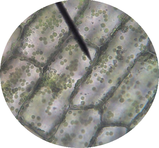
Lab manual exercise 1 lab manual exercise 1 elodea plant cells at 40x 100x 400x with pond water you chloroplast movement in elodea a form of pond weed rosliston astronomy group blog. Students know the characteristics that distinguish plant cells from animal cells, including chloroplasts and cell walls. Students will compare both types of cells. Lab manual exercise 1 lab manual exercise 1 virtual biology labs study botany lab practical flashcards. Leaf structure under the microscope. Scientific investigation 20 compound microscope drawing: Place the leaf or algae sample on a slide and add. Plants have complex cells filled with organelles such as a nucleus, mitochondria, and other structures common to eukaryotes.some plant cells have organelles called chloroplasts that make them green and able to capture energy from light. Label the cells as they appear under high power. Sketch the onion peel cell as seen under the microscope label the. Elodea under dissecting microscope science pics things under. Fresh, green leaf from an elodea plant or take a small sample of algae from the pond water provided. The elodea leaf is composed of two layers of cells.
Plasmolysis is the shrinking of the cytoplasm of a plant cell in response to diffusion of water out of the cell and into a high salt concentration solution. Single cell c4 photosynthesis in aquatic and terrestrial plants a. Two cells will be observed, one from the skin of an onion, and the other from a common aquarium water plant (anacharis). Onion epidermis with large cells under light microscope royalty free stock photo chloroplasts in plant cells 12 animal and plant cells plant cell under a microscope picture. The cell membrane is not visible on the elodea leaf because of its proximity to the much thicker cell wall.
Elodea Leaf Cell Under Microscope Plant Cell Cells Worksheet Lab Activities from i.pinimg.com Plants have complex cells filled with organelles such as a nucleus, mitochondria, and other structures common to eukaryotes.some plant cells have organelles called chloroplasts that make them green and able to capture energy from light. Whats people lookup in this blog: Students know cells function similarly in all living organisms. Observation of plasmolysis when the leaf is flooded with 6%. Hydrilla verticillatea leaf under the microscope hydrilla (esthwaite waterweed, waterthyme or hydrilla) is a genus of aquatic plant that is usually treated as containing only one species: Magnifying and observing cells bioed online. Although some botanists divide this category into several species. Relationship between molecular size solute permeability and the movement of water.
To observe cells with chloroplasts.
They will see cell walls and chloroplasts. Plant cells maintain their normal size. Label the cells as they appear under high power. View a prepared slide of elodea (anacharis), which is an aquarium plant. Add a drop of salt solution (hypertonic solution) to the side of the coverslip and observe the cell. Students know cells function similarly in all living organisms. As the slide warms from the light of the microscope, you may see the chloroplasts moving, a process called cytoplasmic streaming. Microscopic view of canadian waterweed leaf stock photo download. Observe your elodea under the microscope and draw a set of four cells with as many parts labeled as you can identify. Observation of plasmolysis when the leaf is flooded with 6%. Only one layer of cells is in focus when using the high. Label the magnification under which the plant cells are being observed (40x or 100x). Elodea leaf cell illustration from a microscope slide a drop of 10.
The plant is native to the cool and warm waters of the old world in. Solution) and a coverslip and observe the chloroplasts (green structures) and the cell walls. Add a drop of water (hypotonic solution) and a coverslip and observe the chloroplasts (green structures) and the cell walls. Microscopic view of canadian waterweed leaf stock photo download. Pick off an entire healthy looking elodea leaf, with fingers or small scissors and place it on the microscope slide.
Plasmolysis In Elodea Plant Cells Science Netlinks from sciencenetlinks.com During plasmolysis, the cell membrane pulls away from the cell wall. Whats people lookup in this blog: Pass out the elodea images,. Lab manual exercise 1 lab manual exercise 1 elodea plant cells at 40x 100x 400x with pond water you chloroplast movement in elodea a form of pond weed rosliston astronomy group blog. Two cells will be observed, one from the skin of an onion, and the other from a common aquarium water plant (anacharis). Leaf of pondweed aquatic plant stock footage video 100. Place the leaf or algae sample on a slide and add. Rigid walls typically made of cellulose surround plant cells.
Plant cells maintain their normal size.
Elodea under dissecting microscope science pics things under. During plasmolysis, the cell membrane pulls away from the cell wall. Some of these structures can be clearly seen under a compound microscope. Pond water was mixed with the leaf sample so there are some organisms interacting with the lea. Plants have complex cells filled with organelles such as a nucleus, mitochondria, and other structures common to eukaryotes.some plant cells have organelles called chloroplasts that make them green and able to capture energy from light. Only one layer of cells is in focus when using the high. View a prepared slide of elodea (anacharis), which is an aquarium plant. Pass out the elodea images,. Students will compare both types of cells. Label the sketches to note the cell structures that you can identify. Pick off an entire healthy looking elodea leaf, with fingers or small scissors and place it on the microscope slide. Label the magnification under which the plant cells are being observed (40x or 100x). In order to view the membrane, you will add salt to the elodea.
Several types of white cells are visible, including a lymphocyte (b) and some polymorphonuclear leukocytes (labeled c) plant cell under microscope labeled. And see the cell walls clearly.
Share :
Post a Comment
for "Elodea Plant Cell Under Microscope Labeled : Lab Using A Microscope - Pass out the elodea images,."


Post a Comment for "Elodea Plant Cell Under Microscope Labeled : Lab Using A Microscope - Pass out the elodea images,."